Skeletal System Diagram — free printable diagram
Free science resource for teachers · CC BY-NC 4.0
About this illustration
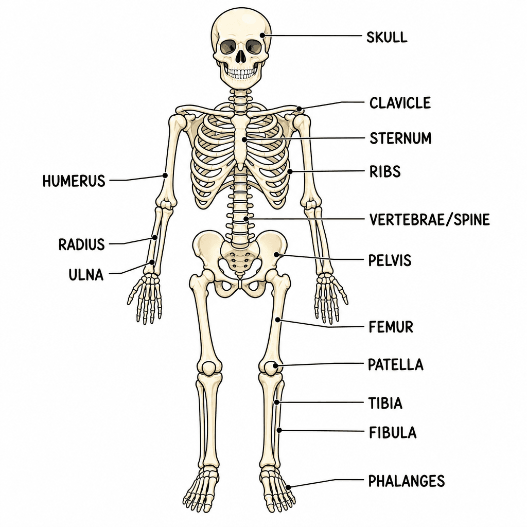
The image presents a full, anterior view of a human skeleton, clearly illustrating the structure of the human body. Various major bones are labeled with connecting lines, including the Skull, Clavicle, Sternum, Ribs, Vertebrae/Spine, Humerus, Radius, Ulna, Pelvis, Femur, Patella, Tibia, Fibula, and Phalanges. This diagram teaches fundamental human anatomy and the names of key bones in the skeletal system. There are no specific cultural or locale cues present, making it universally applicable for biology education. This resource is ideal for slides, reference charts, or as a basis for labeling exercises in worksheets for K-12 science classes. The visual style is a clean, bright, educational illustration with subtle shading that gives the bones a three-dimensional appearance.
How to use
- 1Right-click the image and choose “Save image as”, or use the download button.
- 2Use it in your classroom worksheets, slides or printables — free under CC BY-NC 4.0.
- 3Attribute as “Image by Kuraplan” or link back to kuraplan.com. Not for commercial resale.
Make worksheets with images like this
Kuraplan's editor has the full image library built in — drag-and-drop into a worksheet in seconds.
Browse by subject
15 subjects · 1,990 free illustrations

Cross-Curricular
576 free illustrations
Geography
341 free illustrations
English
316 free illustrations

Maths
189 free illustrations

Religious Education
134 free illustrations
Music
108 free illustrations

Health
107 free illustrations

Art
59 free illustrations

Drama
58 free illustrations

History
45 free illustrations

social_studies
21 free illustrations

tech
16 free illustrations

pe
14 free illustrations

te_reo_maori
5 free illustrations

languages
1 free illustrations