Lungs (unlabeled) — free printable diagram
Free science resource for teachers · CC BY-NC 4.0
About this illustration
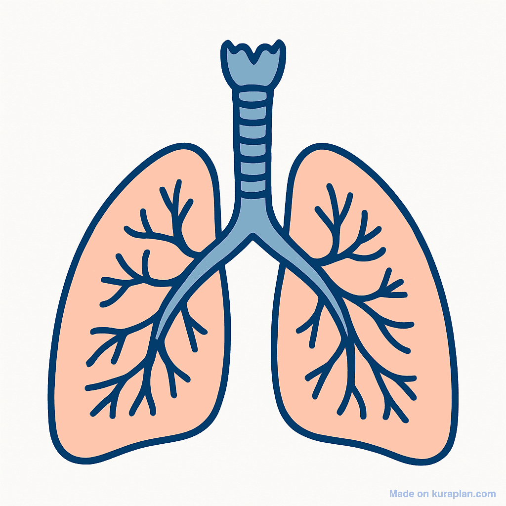
A simplified diagram of the human respiratory system showing two lungs with branching bronchi, trachea connecting upward. Unlabelled.
What to label on a respiratory system diagram
A blank respiratory system diagram is normally used as a labelling exercise. The parts below trace the path air actually takes on the way in — nose and mouth, down the trachea, into the bronchi, along the bronchioles and finally to the alveoli, where gases are exchanged with the blood. Labelling them in that order is usually easier than labelling them by position on the page.
- Nasal cavity
- Warms, moistens and filters incoming air
- Trachea
- The windpipe — held open by rings of cartilage
- Bronchi
- The two large tubes branching into the left and right lung
- Bronchioles
- Progressively smaller tubes inside each lung
- Alveoli
- Tiny air sacs where gas exchange happens
- Lungs
- The pair of organs housing the airways and alveoli
- Diaphragm
- Sheet of muscle below the lungs that drives breathing
- Ribs
- Protect the lungs and move to change chest volume
- Gas exchange happens only in the alveoli. Everything above them — trachea, bronchi, bronchioles — is plumbing that carries air to and from those sacs, which is a distinction students often miss.
- Oxygen passes from the air in the alveoli into the blood, and carbon dioxide passes the other way. Both move by diffusion, from where they are more concentrated to where they are less.
- The alveoli are numerous, thin-walled and richly supplied with blood vessels. Together those three features give a very large surface area and a very short distance to cross, which is what makes the exchange efficient.
- Breathing in is an active movement: the diaphragm contracts and flattens, the ribs move up and out, the chest volume increases and air is drawn in. Breathing out is largely the reverse and is mostly passive at rest.
- The left lung is drawn slightly smaller than the right because the heart occupies space on the left. Students labelling from a diagram often assume the two are identical.
- Breathing and respiration are not the same thing, and the diagram is a good place to make the point: breathing moves air, while respiration is the chemical reaction releasing energy inside cells.
How to use
- 1Right-click the image and choose “Save image as”, or use the download button.
- 2Use it in your classroom worksheets, slides or printables — free under CC BY-NC 4.0.
- 3Attribute as “Image by Kuraplan” or link back to kuraplan.com. Not for commercial resale.
Turn this image into a worksheet
This illustration is already in Kuraplan's editor — describe the worksheet you need and the AI builds it around the image in seconds.
Related illustrations
More free science illustrations
Free worksheets on Lungs (unlabeled)
All free worksheetsBrowse by subject
18 subjects · 4,850 free illustrations
Maths
1,894 free illustrations
Cross-Curricular
835 free illustrations
English
612 free illustrations
Geography
549 free illustrations
Health
200 free illustrations
Social Studies
177 free illustrations
Religious Education
139 free illustrations
Music
128 free illustrations
Art
66 free illustrations
Drama
56 free illustrations
Social Sciences
48 free illustrations
History
47 free illustrations
Arts
26 free illustrations
PE
25 free illustrations
Te Reo Māori
24 free illustrations
Technology
16 free illustrations
Culture
7 free illustrations
Languages
1 free illustrations