Integumentary System Diagram (Skin Cross-section) — free printable diagram
Free science resource for teachers · CC BY-NC 4.0
About this illustration
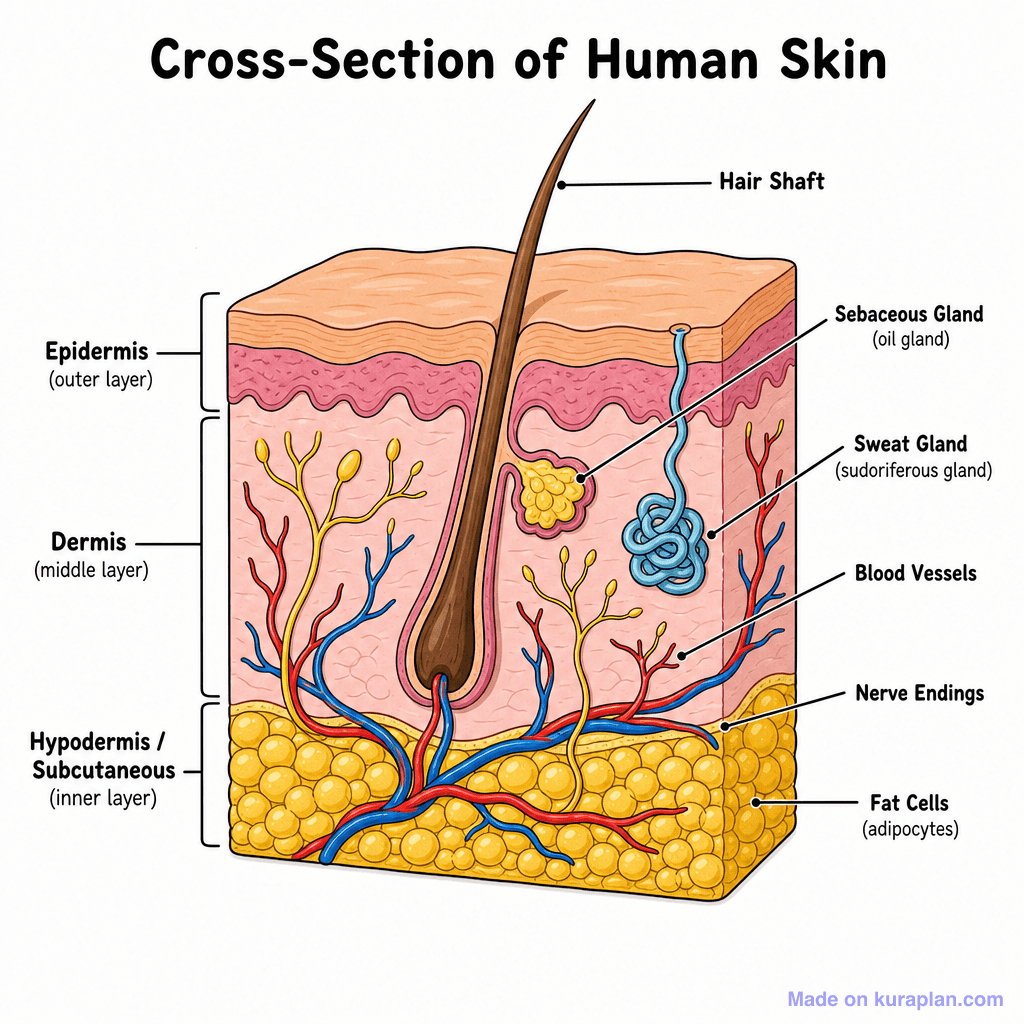
This image displays a labeled cross-section of human skin, showcasing its three primary layers: Epidermis (outer layer), Dermis (middle layer), and Hypodermis/Subcutaneous (inner layer). Within these layers, several key structures are depicted, including a hair shaft, sebaceous gland (oil gland), sweat gland (sudoriferous gland), blood vessels, nerve endings, and fat cells (adipocytes). Each component is clearly identified with text labels and connecting lines. It is designed to teach students about the intricate anatomy and functions of the human integumentary system, suitable for science lessons in K-12 classrooms. This visual is ideal for use on biology worksheets, science lesson slides, or as part of a label exercise. The image utilizes a clear, flat illustration style with high contrast to effectively convey complex biological information.
How to use
- 1Right-click the image and choose “Save image as”, or use the download button.
- 2Use it in your classroom worksheets, slides or printables — free under CC BY-NC 4.0.
- 3Attribute as “Image by Kuraplan” or link back to kuraplan.com. Not for commercial resale.
Turn this image into a worksheet
This illustration is already in Kuraplan's editor — describe the worksheet you need and the AI builds it around the image in seconds.
Related illustrations
More free science illustrations
Free worksheets on Integumentary System Diagram (Skin Cross-section)
All free worksheetsBrowse by subject
18 subjects · 4,850 free illustrations
Maths
1,894 free illustrations
Cross-Curricular
835 free illustrations
English
612 free illustrations
Geography
549 free illustrations
Health
200 free illustrations
Social Studies
177 free illustrations
Religious Education
139 free illustrations
Music
128 free illustrations
Art
66 free illustrations
Drama
56 free illustrations
Social Sciences
48 free illustrations
History
47 free illustrations
Arts
26 free illustrations
PE
25 free illustrations
Te Reo Māori
24 free illustrations
Technology
16 free illustrations
Culture
7 free illustrations
Languages
1 free illustrations