Nervous System Diagram — free printable diagram
Free science resource for teachers · CC BY-NC 4.0
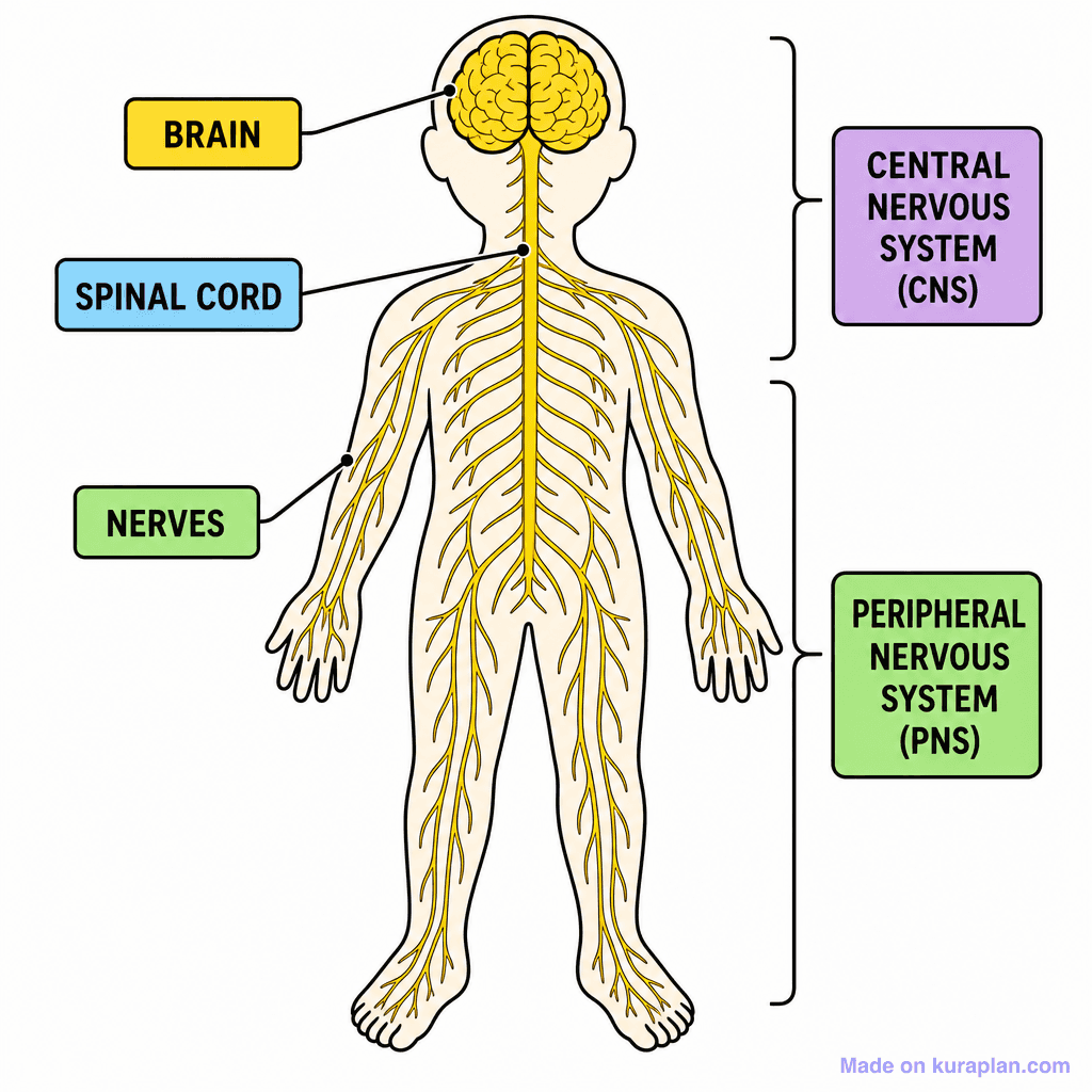
About this illustration
This image depicts a simplified, front-facing, full-body outline of a human, with its nervous system highlighted in yellow. The diagram clearly labels the 'BRAIN', 'SPINAL CORD', and 'NERVES' using colored text boxes and connecting lines. Brackets on the right side further categorize these components into the 'CENTRAL NERVOUS SYSTEM (CNS)' and 'PERIPHERAL NERVOUS SYSTEM (PNS)'. This educational illustration is ideal for teaching elementary and middle school students about human anatomy, specifically the nervous system, and suits classroom activities like worksheets, slides, or basic identification exercises. The visual style is a flat, high-contrast illustration, presented as a clear diagram for learning.
What a nervous system diagram shows
The nervous system carries information around the body as electrical impulses, and it is divided into two parts. The central nervous system is the brain and the spinal cord, where information is processed; the peripheral nervous system is the network of nerves branching out from the spinal cord to every other part of the body. A full-body diagram is drawn to show that branching reach.
- Two main divisions
- Central nervous system (CNS) and peripheral nervous system (PNS)
- Central nervous system
- The brain and the spinal cord
- Peripheral nervous system
- All the nerves branching to the rest of the body
- Brain
- Processes information and coordinates the body
- Spinal cord
- Carries impulses between the brain and the body
- Nerves
- Bundles of neurones
- Neurone
- A nerve cell that carries electrical impulses
- Receptors
- Detect a stimulus — in the eyes, ears, skin and elsewhere
- Effectors
- Muscles and glands that carry out the response
- The pathway is worth learning as a sequence: stimulus, receptor, sensory neurone, central nervous system, motor neurone, effector, response. Every example in this topic is a version of that chain.
- Sorting the labelled parts into CNS and PNS is the main exercise the diagram supports: brain and spinal cord in one group, every nerve in the other.
- A reflex is fast because it does not wait for the brain. The impulse travels into the spinal cord and straight back out to the muscle, which is why pulling your hand from something hot happens before you feel the pain.
- The central nervous system is the part encased in bone — the brain in the skull, the spinal cord in the vertebrae — which is a useful clue for remembering which parts belong to it.
- Nerves are not single cells. A nerve is a bundle of many neurones, in the same way a cable is a bundle of wires.
- The body has a second coordination system, the hormonal one, and the two are usually contrasted: nervous signals are electrical and very fast but short-lived, hormonal signals are chemical, slower and longer-lasting.
How to use
- 1Right-click the image and choose “Save image as”, or use the download button.
- 2Use it in your classroom worksheets, slides or printables — free under CC BY-NC 4.0.
- 3Attribute as “Image by Kuraplan” or link back to kuraplan.com. Not for commercial resale.
Turn this image into a worksheet
This illustration is already in Kuraplan's editor — describe the worksheet you need and the AI builds it around the image in seconds.
Related illustrations
More free science illustrations


Free worksheets on Nervous System Diagram
All free worksheetsBrowse by subject
21 subjects · 4,853 free illustrations
Maths
1,894 free illustrations
Cross-Curricular
835 free illustrations
English
612 free illustrations
Geography
549 free illustrations
Health
200 free illustrations
Social Studies
177 free illustrations
Religious Education
139 free illustrations
Music
128 free illustrations
Art
66 free illustrations
Drama
56 free illustrations
Social Sciences
48 free illustrations
History
47 free illustrations
Arts
26 free illustrations
PE
25 free illustrations
Te Reo Māori
24 free illustrations
Technology
16 free illustrations
Culture
7 free illustrations
Business
1 free illustrations
English
1 free illustrations
English (ELA)
1 free illustrations
Languages
1 free illustrations